Valuable Insights | Common Protein Detection Methods Used in the Laboratory

Extraordinary laboratory use method for determination of protein concentration this one is enough!
Choose the right method, the efficiency of the experiment is doubled!
Part 1 Ultraviolet absorption method (A280 method)
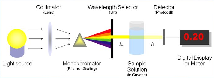
the tryptophan (Trp), tyrosine (Tyr) and phenylalanine (Phe) residues in the protein have UV absorption peaks around 280nm.
By measuring the absorbance value of the protein solution at 280 nm, the protein concentration can be estimated. Based on the Beer-Lambert law: A = εbc,
where A is the absorbance, ε is the molar extinction coefficient, B is the optical path, and c is the concentration. For proteins, ε and B can be treated as constants under certain conditions,
the absorbance A is therefore proportional to the concentration c. FIG. It is generally assumed that the average extinction coefficient ε of a protein is about 1.0(mg/mL)^-1cm ^-1.
The advantages of this method
quick and easy
the operation is very simple and quick, just use the UV spectrophotometer to directly read the absorbance at 280nm.
non-destructive
the sample can be recovered because the sample has not been chemically modified or consumed during the measurement.
No reagents required
in addition to the buffer, no additional chemical reagents are required.
Suitable for high concentrations of protein
it is also relatively accurate in the high concentration range.
The demerits of this method Disadvantages
more interference
nucleic acids (DNA,RNA) also have a strong UV absorption at 260 nm and a certain absorption at 280nm, which will interfere with the determination of proteins.
Some other small molecules may also absorb at 280 nm, causing the results to be biased.
Low sensitivity
for low concentrations of protein solutions, the absorbance value may be too low and the error is large.
Accuracy is affected by protein composition
the tryptophan and tyrosine contents of different proteins are very different, resulting in their molar extinction coefficients also being very different. estimated using the average extinction coefficient,
accuracy can be affected by the amino acid composition of the protein itself. The error is greater if the protein samples contain abnormally high or low levels of Trp and Tyr.
Pure sample required
the sample should not contain a large number of substances that interfere with UV absorption, such as nucleic acids, certain dyes, etc.
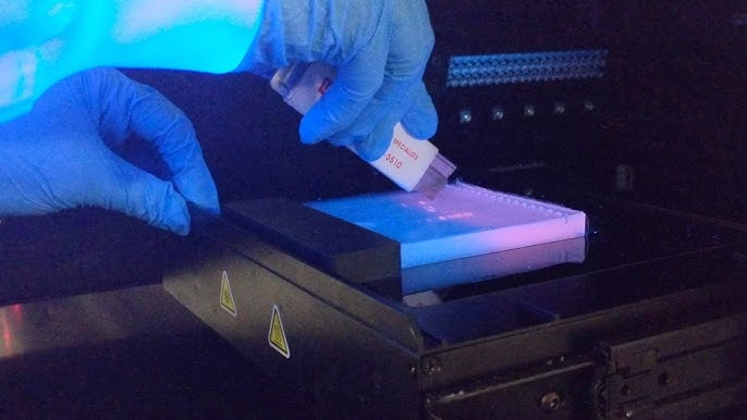
Part 2 Coomassie Brilliant Blue Method (Bradford Method)

coomassie Brilliant Blue G-250 dye binds to the egg autoplasm under acidic conditions, and the dye changes from the red form (maximum absorption wavelength at 465 nm) to the blue form (maximum absorption wavelength at 595 nm).
This color shift is caused by a change in the molecular structure of the dye after the dye binds to the protein. binding is mainly through electrostatic interactions and van der Waals forces,
in particular, the sulfonic acid group of dyes and the non-polarity of basic amino acid residues (such as arginine, lysine and histidine) and aromatic amino acid residues of proteins
regional interactions. The shade of color (absorbance at 595 nm) is directly proportional to the protein concentration.
Merits of this method
high sensitivity
high sensitivity than UV absorption method, suitable for the determination of low concentration of protein.
Easy and quick operation
the reagent is single, the operation steps are few, the reaction is rapid, and it can be completed within a few minutes.
Small interference by salt and buffer in the sample
compared with other colorimetric methods, it is less interfered by common salt and buffer in the sample.
Good stability
the stained blue complex can be stable for a period of time at room temperature, which is convenient to read the data.
demerits of this method Disadvantages
standard curve dependence
it is necessary to use a standard protein of known concentration (usually bovine serum white egg from BSA) to make a standard curve in order to quantify.
The binding ability of different proteins to dyes is different, so the BSA standard curve is used to determine other proteins, and the results may be biased.
protein species dependence
different proteins have different binding capacity to dyes, resulting in different accuracy of quantitative results for different proteins.
The results of protein determination with high content of basic amino acids and aromatic amino acids are relatively accurate, otherwise, the deviation may be large.
The reagent will stain the cuvette
coomassie Brilliant Blue dye easily stains glass or plastic cuvettes and requires careful handling or use of disposable cuvettes.
limited linear range
at higher protein concentrations, saturation of the reaction may occur, resulting in a non-linear standard curve.
Sensitive to detergent
some detergents, such as SDS, can interfere with the Bradford reaction.
Part 3 Lowry method
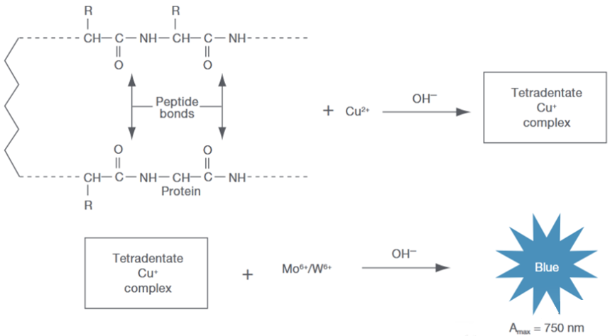
the Lowry method is a classical protein quantification method based on two consecutive chemical reactions. The first step is the peptide bond in the protein and the copper ion.
Under alkaline conditions, the complex is formed, and copper ions are reduced to cuprous ions. The second step is the reduction of the FolinCiocalteu reagent by the reduced cuprous ion
(a mixture of phosphomolybdic acid and phosphotungstic acid), which causes the reagent to develop a blue color. Shades of color (absorbance is usually measured at 750nm or 660nm)
proportional to protein concentration.
Merits of this method
high sensitivity
it is more sensitive than ultraviolet absorption method and Bradford method, and can detect protein at lower concentration.
relatively less affected by protein composition
because it is based on peptide bond reaction, it is less affected by the amino acid composition of different proteins than the Bradford method.
demerits of this method Disadvantages
operation steps cumbersome
multiple reagents are required, and the operation steps are many and time-consuming.
Reagent instability
Folin-Ciocalteu reagents are unstable and easily contaminated by reducing substances, and need to be prepared before use.
More interference
susceptible to interference from a variety of chemicals, including some buffer components (e. g., Tris,EDTA),
and non-ionic detergents, reducing agents, etc. Strict control of sample and reagent purity is required.
time-dependent
color reaction requires precise control of reaction time and temperature, too long or too short reaction time will affect the accuracy of the results.
standard curve dependence
standard curves also need to be made using standard proteins.
Part 4 BCA Method

the principle of the BCA method is similar to that of the Lowry method and is also based on a two-step reaction. The first step is the peptide bond in the protein to the copper ion.
It is reduced to cuprous ions under alkaline conditions. The second step is the reaction of cuprous ions with the BCA reagent (diquinolinic acid) to form a purple BCA-Cu * complex,
the complex has an absorption maximum at 562nm. The depth of color is directly proportional to the protein concentration.
Merits of this method
high sensitivity
the sensitivity of the Lowry method is comparable to that of the Bradford method and the UV absorption method.
operation is relatively simple
the reagent is relatively stable, usually the reagent is provided in a premixed form, and the operation steps are simpler than the Lowry method.
Small interference by detergent
compared with the Lowry method, it is less interfered by some detergents (such as SDS,Triton X-100) and is more suitable for samples containing detergents.
Wide linear range
the linear range of the BCA method is wider than that of the Bradford method.
Good stability
the complex after color development is more stable than the Bradford method, and can be placed for a long time to read the data.
Part5 related consumables recommended
in all aspects of laboratory protein detection, the quality of experimental consumables has an important impact on the reliability of test results.
Aizin Creature rooted in the field of life sciences, with years of accumulated technology and continuous innovation, we have a series of consumables suitable for protein detection,
contains ordinary suction head (low absorption), enzyme labeled plate, micro centrifuge tube, conventional centrifuge tube, cell culture dish, culture plate, culture flask, etc.